
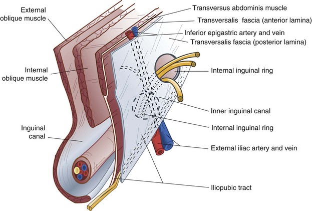
Introduction
- This is an oblique intermuscular passage in the lower part of the anterior abdominal wall , Situated just above the medial half of the inguinal ligament
Location
- Inferior part of the anterolateral abdominal wall
Sponsored Content
 (US/UK)
Length & direction
- It is about 4cm(1.5 inches) long, and is directed downwards, forwards and medially
- The inguinal canal
extends from the deep inguinal ring to the superficial inguinal ring

The Posterior Wall
1.In its whole extent
a. The fascia transversalis
b. The extra peritoneal tissue
c. The parietal peritoneum.
2.In its medial two-thirds
a. The conjoint tendon
b. At its medial end by the reflected part of the inguinal ligament.

Roof of Inguinal Canal
- It is formed by the arched fibres of the internal oblique and transverse abdominis muscles.
Floor of the inguinal canal
- The floor is formed by an incurving of the inguinal ligament, which is part of the external oblique muscle, forming a gutter. (Medially it forms the lacunar ligament).
Deep inguinal ring
- An oval opening in the fascia transversalis situated 1.2 cm above the midinguinal point, and immediately lateral to the stem of the inferior epigastric artery
Superficial inguinal ring
- Is a triangular gap in the external oblique aponeurosis .
- It is shaped like an obtuse angled triangle .
- The base of the triangle is formed by the pubic crest, the two sides of the triangle from the lateral or lower and the medial or upper margins of the opening.
- It is 2.5 cm long and 1.2 cm broad at the base these margins are referred as crura.
Anterior Relations of Inguinal Canal
- In its whole extent
- Skin
- Superficial fascia
- External oblique aponeurosis
- In its lateral one-third
- The fleshy fibres of the internal oblique muscle.
Structures Passing Through Inguinal Canal
- The spermatic cord in males, or the round ligament of the uterus in females, enters the inguinal canal through the deep inguinal ring and passes out through the superficial inguinal ring.
- The ilioinguinal nerve enters the canal through the interval between the external and internal oblique muscles and passes out through the superficial inguinal ring.
Constituents of Spermatic Cord
- Vas Deferens
- Arteries
- Testicular Artery
- Cremasteric Artery
- Artery of Vas Deferens
- Pampiniform plexus of veins
- Lymph Vessels from Testis
- Genital Branch of Genitofemoral Nerve
- Sympathetic Nerve Plexus around Artery of Vas Deferens
Coverings of Spermatic Cord
From within outwards, these are as follows:
1.The internal spermatic fascia , derived from the fascia transversalis; it covers the cord in its whole extent .
2.The cremasteric fascia is made up of the muscle loops constituting the cremaster muscle, and the intervening areolar tissue. It is derived from the internal oblique and transversus abdominis muscles.
Mechanisms Preventing Inguinal Hernia
Obliquity of the inguinal canal:The two inguinal rings do not lie opposite to each other. Therefore, when the intra-abdominal pressure rises the anterior and posterior walls of the canal are approximated, thus obliterating the passage. This is known as the flap valve mechanism.
The superficial inguinal ring is guarded from behind by the conjoint tendon and by the reflected part of the inguinal canal.
The deep inguinal ring is guarded from the front by the fleshy fibres of the internal oblique.
Shutter mechanism of the internal oblique: This muscle has a triple relation to the inguinal canal. It forms the anterior wall, the roof, and the posterior wall of the canal. When it contracts the roof is approximated to the floor, like a shutter.
Ball valve mechanism of Cremaster: Contraction of the cremaster helps the spermatic cord to plug the superficial inguinal ring
Slit valve mechanism of Superficial Ring: Contraction of the external oblique results in approximation of the two crura of the superficial inguinal ring . The integrity of the superficial inguinal ring is greatly increased by the intercrural fibres.
Hormones: They may play a role in maintaining the tone of the inguinal musculature
Hesselbach's Triangle
Hesselbach’s (Inguinal) Triangle is an important structure as it is the site for direct hernias. The triangle has the following borders:
- Medial border of rectus abdominus(medially)
- Inguinal ligament (inferiorly)
- Inferior epigastric vessels(laterally)

Laparoscopic Inguinal Hernia Repair- Surgical Anatomy
Myopectineal orifice of Fruchaud
- It is a potential weak area in the lower abdominal wall through which groin hernias occur. It is bounded:
- Above by conjoint tendon
- Below by the pectineal line of the superior pubic ramus
- Laterally by the iliopsoas muscle
- Medially by the lateral border of the rectus muscle.
- This serves as the passage for blood vessels, nerves, lymphatics, muscles and tendons between the abdomen and the lower limb.
- This is the site for direct, indirect and femoral hernias.
- All the three can be repaired by a single piece of mesh by covering this orifice
Space of Retzius and Bogros
Related posts:
LAPAROSCOPIC EXTRAPERITONEAL INGUINAL HERNIA REPAIR IS USUALLY PERFORMED IN WHICH OF THE FOLLOWING SPACE?
http://www.worldsurgeryforum.net/2016/05/laparoscopic-extraperitoneal-inguinal.html
https://www.worldsurgeryforum.net/2017/07/surgical-anatomy-of-inguinal-canal.html
SURGICAL ANATOMY OF INGUINAL CANAL
https://www.worldsurgeryforum.net/2017/07/surgical-anatomy-of-inguinal-canal.html


