| 1 | Thyroid Status Examination - OSCE Guide |  | 2015-02-25 15:22:54 | ziaYBkgEZNU | 00:06:29 |
| 2 | Hip examination for OSCE revision |  | 2012-02-22 14:41:26 | ROzFs4FQnTQ | 00:07:31 |
| 3 | Hip Joint Examination - OSCE Guide |  | 2015-08-19 18:56:16 | KAgRJTsXHrU | 00:06:01 |
| 4 | Clinical Examination - Head and Neck Lymph nodes |  | 2015-10-08 09:15:32 | _N89T_Yqu68 | 00:02:53 |
| 5 | Shoulder Examination - OSCE Guide (New Version) | 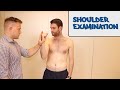 | 2015-08-24 22:52:00 | TDs1IOFYnMo | 00:06:37 |
| 6 | Spine Examination - OSCE Guide (New Version) |  | 2015-08-03 06:57:21 | CJVGpciEMoI | 00:04:59 |
| 7 | Examination Of Parotid Gland |  | 2011-11-06 13:17:51 | Gx1vVwA5AFU | 00:02:54 |
| 8 | Tests For Examination Of The Lower Back - Everything You Need To Know - Dr. Nabil Ebraheim | 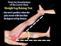 | 2013-03-07 22:36:40 | zfWKcTixM6E | 00:06:02 |
| 9 | Lachman's Test , ACL Injury - Everything You Need To Know - Dr. Nabil Ebraheim |  | 2015-07-17 21:30:01 | tf2xBJZFcg4 | 00:03:12 |
| 10 | ISBAR - Case Study 1 |  | 2012-03-23 02:18:22 | 1Wl9qogPw1E | 00:04:28 |
| 11 | ISBAR - Case Study 2 |  | 2012-03-23 02:18:22 | AmZKJ3JAPsE | 00:04:03 |
| 12 | Elbow Examination | 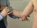 | 2007-11-19 07:43:47 | bMJjbIT3zek | 00:01:52 |
| 13 | Breaking bad news |  | 2016-06-30 07:58:10 | k0_04r8hPAs | 00:05:55 |
| 14 | How to Break Bad News.avi |  | 2012-01-14 20:40:10 | hkYCcFgSEZc | 00:08:33 |
| 15 | electrosurgery principles |  | 2011-02-06 18:13:52 | 7LW78yoaEe0 | 00:11:38 |
| 16 | How to Palpate the Salivary Glands |  | 2014-11-19 04:35:48 | ryQh39mA6Hg | 00:01:57 |
| 17 | Qpercom presents OMIS in Action: Breaking Bad News OSCE Station |  | 2013-05-23 15:08:29 | QvW_g6w8uY0 | 00:09:08 |
| 18 | Breast Examination - OSCE Guide |  | 2015-07-23 23:35:35 | 76g_tNWMhCE | 00:05:40 |
| 19 | CSA case = Breaking Bad News (Done well) |  | 2013-06-30 18:18:38 | lJN6g0V5Q-U | 00:09:25 |
| 20 | Diabetic foot examination - OSCE guide (New Version) |  | 2015-03-27 15:24:29 | vwIyulPnXcg | 00:05:32 |
| 21 | Gait Assessment - Normal Gait and Common Abnormal Gaits |  | 2015-02-19 21:51:58 | PZBiv0uSXVg | 00:23:14 |
| 22 | Peripheral Vascular Examination - OSCE Guide (New Version) |  | 2015-04-21 11:23:18 | 6beOTEKx1ek | 00:06:25 |
| 23 | Abdominal Examination - OSCE Guide (New Version) |  | 2015-02-19 15:24:25 | XOefpxm38bc | 00:06:57 |
| 24 | MENTOR CSA CONSULTS - ANGRY PATIENT |  | 2013-08-09 13:36:24 | POiRQrrQJQU | 00:07:58 |
| 25 | Varicose Vein Examination for Finals |  | 2008-05-27 17:54:32 | jsCoSHP4RmE | 00:09:20 |
| 26 | Laparoscopic Anatomy - WITH labels |  | 2013-10-19 20:32:07 | h3mUMhtIZ_A | 00:04:45 |
| 27 | Differential diagnosis of abdominal pain according to abdominal regions |  | 2013-03-05 10:21:48 | HbG7t7vfRRM | 00:12:24 |
| 28 | Deleted video |  | 2016-02-09 14:25:07 | 2cyA3zHqq2s | 00:16:26 |
| 29 | Diagnosis & Examination of the floor of the mouth(Bimanual technique)(Dentistry) |  | 2013-04-20 11:42:55 | FPeWSkBSoOo | 00:00:28 |
| 30 | MCC Extraoral Exam |  | 2014-09-18 14:17:57 | K3Gx6GHn0OE | 00:12:52 |
| 31 | SKELETAL SYSTEM ANATOMY: Cranial fossa of the human skull | 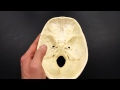 | 2013-03-06 22:00:56 | weJ-eaDRIGs | 00:06:38 |
| 32 | SKELETAL SYSTEM ANATOMY: Anterior aspect of the human skull |  | 2013-03-06 22:19:25 | We2QFap4qyU | 00:05:14 |
| 33 | SKELETAL SYSTEM ANATOMY: Lateral aspect of the human skull | 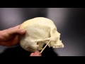 | 2013-03-06 22:05:39 | XLDiuHE3ToU | 00:03:55 |
| 34 | SKELETAL SYSTEM ANATOMY: Inferior aspect of the human skull |  | 2013-03-06 21:45:15 | cUnIgkZ10KY | 00:05:42 |
| 35 | TMJ Video - Temporomandibular Joint | 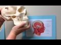 | 2014-04-29 04:52:49 | 4vQwyMHPXkk | 00:06:49 |
| 36 | Vascular OSCE Examination | 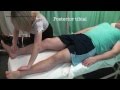 | 2013-01-19 22:00:34 | 5Y-0OWZDbeo | 00:03:37 |
| 37 | How to talk about the normal abdominal plain X-ray |  | 2014-07-28 11:40:04 | iVCicL158BY | 00:13:34 |
| 38 | Radial Nerve Palsy, injury - WRIST DROP . Everything You Need To Know - Dr. Nabil Ebraheim |  | 2010-11-09 15:19:52 | _Cu6ttAhe8Y | 00:04:54 |
| 39 | Chest Tube ATLS |  | 2014-06-13 12:12:33 | qR3VcueqBgc | 00:03:17 |
| 40 | Chest Tube Insertion NEJM |  | 2014-01-10 05:44:56 | f_JWzLiUNhE | 00:16:07 |
| 41 | Joseph Musowoya - Trialling a patient assessment checklist for critically ill surgical patients |  | 2014-11-25 19:20:46 | jggurLNJYRM | 00:08:11 |
| 42 | Clinical Brain Death Testing |  | 2015-04-13 02:12:30 | qiZBGFWvv4E | 00:18:52 |
| 43 | The Effect of Supplemental Oxygen (ABG Interpretation - Lesson 17) | 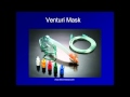 | 2012-05-31 07:25:26 | 9klrDCall9M | 00:28:36 |
| 44 | 6 Easy Steps to ABG Analysis |  | 2013-05-17 16:48:14 | WUf-cPpnrXw | 00:13:19 |
| 45 | Arterial blood gas interpretation in 5 min |  | 2013-10-16 10:27:49 | Y0Q4inCKv2A | 00:05:00 |
| 46 | Arterial Puncture for Blood Gas Analysis.flv |  | 2011-11-24 16:49:17 | YuFK22n-tvI | 00:09:07 |
| 47 | Initial Assessment of a Trauma Patient - Normal Scenario.wmv |  | 2012-02-17 15:28:36 | hLuC0T7RsKI | 00:14:42 |
| 48 | Initial Assessment of a Trauma Patient - Multi-System Injury (Part 1).wmv |  | 2012-02-17 15:20:35 | n5DUC1Lit08 | 00:11:42 |
| 49 | Reading the 12-lead ECG/EKG - six quick steps |  | 2013-07-15 09:53:33 | lhrajncAh9A | 00:13:23 |
| 50 | Intro to EKG Interpretation - The 5 Cardinal Rules |  | 2014-03-15 07:12:53 | r2zQvatebxA | 00:11:58 |
| 51 | Gross Anatomy of Pancreas |  | 2015-04-25 15:22:40 | 1oj13Uua00A | 00:03:05 |
| 52 | SKELETAL SYSTEM ANATOMY: Bones of the thigh- Femur |  | 2013-03-04 18:47:14 | Xb-U3Jjj-n4 | 00:03:26 |
| 53 | The Facial Nerve (CNVII): Animated Review |  | 2016-04-26 15:09:22 | AMAR-hv7knI | 00:06:37 |
| 54 | How to interpret an abdominal CT |  | 2015-11-23 13:52:25 | Eg3GeUmOeYE | 00:25:18 |
| 55 | Catheterisation - OSCE Guide |  | 2015-05-02 18:54:25 | 2iLPfCAMgZs | 00:04:34 |
| 56 | Basic life support - Head tilt Chin lift Jaw thrust |  | 2015-03-29 07:52:57 | XNZed1oBeTA | 00:02:25 |
| 57 | How to perform Tracheostomy - An Emergency Procedure - Operative Live Surgery by Dr Vaidya |  | 2013-02-26 14:09:49 | 9EOUeWIz5yM | 00:02:31 |
| 58 | tracheostomy |  | 2011-12-31 10:54:48 | Qc0tNXF8tU8 | 00:05:36 |
| 59 | Centeral venous cath. (internal jugular vein).wmv1 |  | 2010-06-16 19:25:22 | 4-uyTUzPSJ8 | 00:07:31 |
| 60 | Centeral venous cath.(internal jugular vein).wmv2 |  | 2010-06-16 19:51:49 | UrHwKQY8qEQ | 00:08:00 |
| 61 | femoral hernia repair |  | 2016-03-09 22:23:43 | 1UnfSdPA59Q | 00:14:13 |
| 62 | How open femoral hernia surgery is carried out |  | 2013-08-14 13:24:59 | dy2s6BbohBs | 00:02:00 |
| 63 | Laparoscopic Splenectomy : Minimally invasive spleen removal surgery |  | 2015-11-09 09:31:18 | zrB4i32ORhs | 00:11:54 |
| 64 | Two Incision Four Compartment Fasciotomy |  | 2010-01-18 21:48:25 | -1NDJkFH1vM | 00:04:15 |
| 65 | Trendelenburg Test (CR) |  | 2011-08-14 04:46:04 | mA3enWJ53Kc | 00:00:36 |
| 66 | Froment's Sign |  | 2012-08-20 19:15:54 | yJTIhm1VfSI | 00:01:14 |
| 67 | Tinel Sign: Wrist⎟Carpal Tunnel Syndrome |  | 2015-12-14 18:21:22 | U8cPjPeZgFw | 00:01:59 |
| 68 | Carpal Tunnel Syndrome - Everything You Need To Know - Dr. Nabil Ebraheim |  | 2015-09-25 23:27:45 | tjyU55ilshQ | 00:12:48 |
| 69 | A guide to intravenous fluids (IV) |  | 2014-08-21 19:21:15 | pln-x9YKnK0 | 00:09:46 |
| 70 | 2. Examination of an Ulcer - Clinical Examination - Dr. Vaidya |  | 2016-04-27 10:16:57 | Y6FsRHFcyoI | 00:14:03 |
| 71 | Cardiovascular Examination - OSCE Guide (New Version) |  | 2015-02-23 16:53:40 | eBnzjerIHj0 | 00:07:38 |
| 72 | Excision of skin lesion (simulated) |  | 2014-04-02 16:03:19 | hjtRQygj20Q | 00:01:53 |
| 73 | Naevus Excision |  | 2015-10-01 05:49:54 | TABHPV3XUvU | 00:03:19 |
| 74 | OSCE Guides - Approach to the patient with knee pain |  | 2016-08-27 15:22:49 | 5pixSqEhg9c | 00:17:18 |
| 75 | Dr. Gillard's presentation of Lumbar Spine and Disc Anatomy |  | 2014-08-20 12:50:08 | XXNA87SbuUc | 00:25:52 |
| 76 | Dr. Gillard lectures on How to Read Your Lumbar MRI | 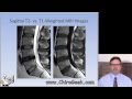 | 2014-08-22 12:52:25 | 3YnMzXOTd2I | 00:23:39 |
| 77 | Chest Anatomy - Trachea, Oesophagus, Lungs and Heart Anatomy - Radiology Pictures and Images |  | 2016-09-11 12:34:28 | 4LBvWLAVdd4 | 00:06:58 |