Showing posts with label neuro. Show all posts


A helmet with infrared light that can heal brain after mild head injury or concussion. This therapy is called Photobiomodulation.

Mild traumatic brain injuries, such as concussions, account for 75% of all cases in the U.S.

Acceleration and deceleration within the cranial vault leads to tearing and stretching of nerve fibers. Alteration of ionic balance and mitochondrial dysfunction makes it difficult for the cells to function while also limiting energy available for healing.

This wearable technology uses light therapy to speed brain recovery. This helmet emits near-infrared light. The light will penetrate the skull and reverse the impaired cellular metabolism that characterizes concussions.

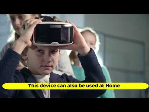
This device can be used in an outpatient setting.

#Concussion
#Traumatic #Head #Injury


Research Paper: https://www.ncbi.nlm.nih.gov/pmc/articles/PMC5927185/
Read More: https://www.concussionalliance.org/light-therapy-photobiomodulation 
Neuro Urology and The Neurogenic Bladder

1. Neurogenic bladder refers to dysfunction of the urinary bladder due to disease of the central nervous system or peripheral nerves involved in the control of micturition . 
2. Non Neurogenic bladder refers to dysfunction of the urinary bladder due to dynamic disturbance of genitourinary system. 
 
Micturition Pathway
3. detrusor muscle of the bladder is innervated by parasympathetic neurons located in the S2-S4  column
 

The CT comma sign is a characteristic sign seen in head trauma. It is the presence of concurrent epidural and subdural hematomas, which gives the characteristic appearance of this sign as a "comma" shape.

The CT comma sign is a characteristic sign seen in head trauma. It is the presence of concurrent epidural and subdural hematomas, which gives the characteristic appearance of this sign as a "comma" shape.

The CT comma sign is a characteristic sign seen in head trauma. It is the presence of concurrent epidural and subdural hematomas, which gives the characteristic appearance of this sign as a "comma" shape.
The CT comma sign is a characteristic sign seen in head trauma. 
It is the presence of concurrent epidural and subdural hematomas, 
which gives the characteristic appearance of this sign as a "comma" shape.
           

70/M 

Chief Complaints: Numbness Lt Arm, 1 Episode of Partial seizure,Lt grip weakness since today morning. 

H/o Fall 3 days back. 

K/C/O HTN/DM/Post CABG 1994 with implanted defibrillator device (CRTD)/ Post Mechanical Thrombectomy for Rt MCA territory Infarct (March2020) on Ecospirin and T. APIXABAN(last dose today). 

Kn chronic  smoker and alcoholic. 

O/E 

E4V5M6
Pupils B/L 2mm RTL . 
Power Lt side 4/5. 
Left Hand grip 70%.

Since the patient was having multiple comorbidities with deranged coagulation and the GCS was 15, it was decided to manage the patient conservatively. Ecospirin and APIXABAN were stopped after consulting with a cardiologist and neurologist. 

#neuroradiology#neuroscience
#neurosurgeon #brainmri #braincat 

Reference


    MRI Spectroscopy : Neurosurgery Notes

    1. MR spectroscopy provides a measure of brain chemistry.
    2. The most common nuclei that are used are 
      • 1H (proton)
      • 23Na (sodium)
      • 31P (phosphorus). 
      • Proton spectroscopy is easier to perform and provides much higher signal-to-noise than either sodium or phosphorus.
    3. MRS can be performed within 10-15 minutes and can be added on to conventional MR imaging protocols. 
    4. It can be used to serially monitor biochemical changes in tumors, stroke, epilepsy, metabolic disorders, infections, and neurodegenerative diseases. 
    5. They require interpretation and should always be correlated with the MR images before making a final diagnosis.
    Hypothalamus Neuroanatomy/ Neurosurgery Notes


     Video Link:Neurosurgery written board crash course - hypothalamus 


    1. hypothalamus as the name suggests is directly underneath the thalamus and it's directly above the pituitary 
    2. borders of the hypothalamus can be drawn by a triangle 
      • line between the anterior commissure and a posterior commissure and it's called the ACPC line
      • line between the anterior commissure and the optic chiasm roughly boarded by the lamina terminalis 
      • line between the chiasm and the posterior commissure 


Q1. The “Nissl substance” represents which organelle of neuron?
A. Golgi complex
B. Nucleolus
C. Rough endoplasmic reticulum
D. Mitochondria

Ans: The cytoplasm of a neuron shows the presence of a granular material that stains intensely with basic dyes called Nissl substance (also called Nissl bodies or granules) . These bodies are rough endoplasmic reticulum

Q2. Which of the following provides myelin sheath to the axons of the CNS?
A. Astrocytes
B. Oligodendrocytes
C. Microglia
D. Ependymocytes

Ans: Oligodendrocytes form myelin sheath in CNS. Schwann cells form myelin sheath in PNS

Q3. The perivascular foot of the “blood–brain barrier” is an extension from the:
A. Oligodendrocyte
B. Ependymocyte
C. Astrocyte
D. Microglia

Ans: Astrocytes form the perivascular feet around a capillary. Astrocytes act as insulators, nourish the neurons, help form blood-brain barrier.

Q4. Sensation of pain is detected by:A. Mechanoreceptor
B. Chemoreceptor
C. Nociceptor
D. Thermoreceptor


Ans: Nociceptor

Q5. The cerebral aqueduct is developed from the cavity of:A. Rhombencephalon
B. Mesencephalon
C. Telencephalon
D. Diencephalon


Ans: The cavity of each telencephalic vesicle becomes the lateral ventricle. The cavity of  iencephalon (along with the central part of the telencephalon) becomes the third ventricle. The cavity of the mesencephalon remains narrow, and forms the cerebral aqueduct (aqueduct of Sylvius). The cavity of the rhombencephalon forms the fourth ventricle. Its continuation in the spinal cord is the
central canal


Q6. The failure of closure of the cranial end of neural tube gives rise to:A. Anencephaly
B. Hydrocephalus
C. Microcephaly
D. Meningomyelocoele


Ans: The neural tube remains open in the region of the brain because of nonclosure of the anterior neuropore. This results in anencephaly. Brain tissue, which is exposed, degenerates

Q7. By which week of intrauterine life does the neural tube close?
A. Fourth
B. Fifth
C. Sixth
D. Seventh


Ans: At 4 weeks the neural tube is normally completely closed (Ref: https://embryology.med.unsw.edu.au/embryology/index.php/Timeline_human_development#Neural)

Q8. The cervical flexure of the neural tube occurs:A. Between the forebrain and midbrain
B. In the midbrain
C. Between hindbrain and spinal cord
D. In the hindbrain
 

Ans: The cervical flexure lies at the junction of the rhombencephalon and the spinal cord  


Q9:Rabies virus, from the site of bite, travels along nerves by 
A. Forward Axoplasmic Flow
B: Reverse Axoplasmic flow
C: Dendritic Flow
D: Along the Endoneurium of nerve fibres


Ans: Rabies virus, from the site of bite, travels along nerves by reverse axoplasmic flow.

Q10:Polio virus is also transported from the gastrointestinal tract through :


A. Forward Axoplasmic Flow
B: Reverse Axoplasmic flow
C: Dendritic Flow
D: Along the Endoneurium of nerve fibres

Ans: Polio virus is also transported from the gastrointestinal tract through reverse axoplasmic flow.


Q11:Tetanus bacteria, in contrast, travels from the site of infection to the brain by:
A. Forward Axoplasmic Flow
B: Reverse Axoplasmic flow
C: Dendritic Flow
D: Along the Endoneurium of nerve fibres

Ans: Tetanus bacteria travels from the site of infection to the brain along the endoneurium of nerve fibres
  
Reference:

History Taking in Neurosurgery : Headache

What are the pain sensitive intracranial structures?

Brain itself is pain insensitive. 

The following intracranial structures are pain-sensitive:
  1. Meningeal arteries
  2. Proximal portions of the cerebral arteries
  3. Dura at the base of the brain
  4. Venous sinuses
  5. Cranial nerves 5, 7, 9, and 10, and cervical nerves 1, 2, and 3

What are the mechanisms causing headaches?

  • Distortion or traction of Dura, Venous Sinuses or Blood vessels: 

    • Drainage of CSF in erect posture causes headache, secondary to traction on the venous sinuses when the brain sinks toward the tentorium as it loses CSF flotation
    • Intracranial mass distorts the dura or the arteries at the base of the brain 
    • Distortion due to raised ICP 

  • Distension of a vessel

    • Distension of extracranial and occasionally intracranial arteries is thought to be the cause of pain in migraine (activate the trigeminal nerve terminals in the vessel wall)

  • Inflammation

    • Inflammation in the subarachnoid space can result in headache. Inflammation can be caused by infection, hemorrhage, or chemical irritation
    • Inflammation of vessel wall by autoimmune process. eg Giant cell arteritis

  • Referral of Pain

    • Lesions above the tentorium - referred pain in trigeminal nerve distribution (the forehead or behind the eye) - because the dura in this region is supplied by the trigeminal nerve
    • Lesions in the posterior fossa
      • referred pain in the ear and the back of the head - because this part of the dura is supplied by cranial nerves 9 and 10 and the upper three cervical roots
      • refer pain to orbit -  termination of orbital (ophthalmic division) pain nerve fibers in the lowest part of the spinal nucleus of the trigeminal nerve, which also receive termination of the upper cervical pain afferent nerve fibers 
      • referred pain to ear - Irritation of cranial nerves 7, 9, and 10 - because the ear has cutaneous supply from each of these nerves as well as cranial nerve 5.

What are the types of Headaches?

For the purpose of history taking, headaches can be divided into two types:
  • Primary- no identifiable cause on examination or investigation and diagnosis is based on  recognizing a pattern, e.g.
    • Migraine
    • Cluster headache
    • Tension-type headache
  • Secondary - definite identifiable cause on examination or investigation, e.g. 
    • Brain tumors
    • Meningitis
    • Sub-arachnoid hemorrhage 
  • 90% - primary headaches, less than 10% are secondary headaches (Rasmussen 1991)

What are the headache "Red Flags"?

  • Worst Headache ever
  • New onset Headache
  • Onset after age of 50 yrs
  • Change in pattern of headache
  • Worsening headache
  • Sudden onset during exertion, sneezing, coughing
  • Headache with postural variation
  • Headache in setting of malignancy or HIV
  • Headache associated with Neurological symptoms or signs
  • Associated with systemic symptoms - fever, weight loss and chronic cough

What history to take in a patient presenting with Headache?


Duration
Exact duration as reported by patient
NEW onset headache or something that has been there since before and has worsened now

Onset ( sudden/ gradual )
Acute onset, severe, first and worst headache, the common possibilities are sub-arachnoid hemorrhage, vascular dissection, pituitary apoplexy
Gradual onset – migraine(mins to days), SDH, GCA(days to months)

Severity
verbal rating scale from 0 to 10

Character
Pulsatile or throbbing or hammering (Raised ICP Headaches/ Migraine)
Dull featureless pain (Tension type headache)
Boring sharp- cluster HA

Time of occurrence
Raised ICP headaches - More in morning, May waken the patient at an early hour

Frequency
Increase in frequency - red flag

Distribution
Frontal or Holocranial - Raised ICP
Band like - tension headaache


Aggravating factors
Exertion, coughing, sneezing, stooping, and straining at stool
Changes in posture (increases in supine - ↑ICP , increases in upright – low CSF pressure headache)

Relieving factors
Improvement on lying flat - low pressure headache

Associated features
Blurring of Vision - Papilloedema
Diplopia, the commonest cause of which is abducens nerve paresis
Nausea & vomiting – migraine, ↑ICP
Neck stiffness – meningeal process
Changes in consciousness
Focal neurological symptoms


References


The BASICS trial provides evidence to support the adoption of antibiotic shunts who are having their first ventriculoperitoneal shunt insertion. 

Summary 

Background 

The aim of this trial was to determine the clinical and cost-effectiveness of antibiotic (rifampicin and clindamycin) or silver shunts compared with standard shunts at reducing infection. 

Methods 

It was a parallel, multicentre, single-blind, randomised controlled trial, including patients with hydrocephalus of any aetiology undergoing insertion of their first ventriculoperitoneal shunt irrespective of age at 21 regional adult and paediatric neurosurgery centres in the UK and Ireland. Patients were randomly assigned (1:1:1 in random permuted blocks of three or six) to receive standard shunts (standard shunt group), antibiotic-impregnated (0·15% clindamycin and 0·054% rifampicin; antibiotic shunt group), or silver-impregnated shunts (silver shunt group) . Followed up for at least 6 months and a maximum of 2 years. The primary outcome was time to shunt failure due the infection and was analysed with Fine and Gray survival regression models for competing risk by intention to treat. 

Findings 


Between June 26, 2013, and Oct 9, 2017, 1605 aged up to 91 years were randomly assigned to receive either a standard shunt (n=536), an antibiotic-impregnated shunt (n=538), or a silver shunt (n=531). 32 (6%) of 533 evaluable patients in the standard shunt group had a shunt revision for infection, compared with 12 (2%) of 535 evaluable patients in the antibiotic shunt group (cause-specific hazard ratio [csHR] 0·38, 97·5% CI 0·18–0·80, p=0·0038) and 31 (6%) of 526 patients in the silver shunt group (0·99, 0·56–1·74, p=0·96). 

Interpretation 

The BASICS trial provides evidence to support the adoption of antibiotic shunts in UK patients who are having their first ventriculoperitoneal shunt insertion 

Diagram of Pupillary Light Reflex Pathway
Pupillary Light Reflex Pathway #neurosurgeon #neuro #neurosurgery #neurological #Pupil #neuroscience #neurology #neurochirurgie #lightreflex #pupillary #Lightreflexpathway https://ift.tt/33tMQAo via WSF Instagram

Anatomy of Basal Ganglia - Axial View

Structures generally included in the basal ganglia are the caudate, putamen, and globus pallidus in the cerebrum, the substantia nigra in the midbrain, and the subthalamic nucleus in the diencephalon.

Basal ganglia's primary function is likely to control and regulate activities of the motor and premotor cortical areas so that voluntary movements can be performed smoothly.

Dysfunction results in a wide range of neurological conditions including disorders of behaviour control and movement.

Those of behaviour include Tourette syndrome, obsessive–compulsive disorder, and addiction.

Movement disorders include, most notably Parkinson's disease, which involves degeneration of the dopamine-producing cells in the substantia nigra, Huntington's disease, which primarily involves damage to the striatum, dystonia, and hemiballismus


 #neurosurgeon #neuro #neurosurgery #neurological #basalganglia #neuroscience #neurology #neurochirurgie #neuroradiology #BGBleed #caudate #caudatenucleus https://ift.tt/33tMQAo via WSF Instagram

Topics