Showing posts with label neurology. Show all posts
Can Gas cause Headaches?

Here is a common query by patients which makes us smile often. Gas causing headaches! 


And you would be surprised that this is a very common query being searched on Google every day.


Have explored the evidence related to it in this brief blog. Your views and comments are welcome. 


If you go through the searches people do on google related to headaches, one very intriguing query that will make you wonder why people have been searching for a correlation between Abdominal Gas and Headaches. 


Is there really a correlation or is it just a piece of misinformation prevalent in the community? Or is it being spread by alternative medicine practitioners carried on with traditional teachings without any scientific evidence? 


Below are the common related keyword searches and the search volumes in India (Source: Ubersuggest):

  • Does gas cause headache - 320 searches per month
  • can gas cause headache - 590 searches per month
  • can gas cause headaches - 260 searches per month
  • can gas cause headaches and dizziness - 90 searches per month
  • does stomach gas cause headache - 30 searches per month
Other related searches
  • can gas problem cause headache
  • why gas causes headache
  • is gas cause headache
  • will gas cause headache
  • headache due to gas during pregnancy
  • how does stomach gas cause headache
  • does gas in stomach cause headache
  • does acidity and gas cause headache
  • can gas cause a headache
  • do gas cause headaches
  • does gas problems cause headaches
  • headache and vomiting due to gas
  • how does gas cause headache
  • can acidity cause headache and vomiting
  • does gas give you headaches
  • does natural gas cause headaches
  • does gas cause migraines
  • can gas cause head pain
  • gas causing headache remedy

What is the direct answer to this popular question?

The answer to this question is NO, the gas cannot directly cause headaches in a normal individual unless you develop air embolism. This is seen in scuba divers when they do a sudden ascent after a deep dive. It can also be seen after head and neck injuries where air can make way into the blood circulation through a bleeding vessel.

However, due to this prevalent belief, there have been many studies that have found that some headaches do get relieved on the treatment of the associated gastric and intestinal disorders (causing gastric discomfort which is perceived as gas).

Gastrointestinal Disorders associated with Headaches

 Gastric and intestinal disorders which cause gastric discomfort (perceived as Gas) and have also been found to have some association to headaches are (1) :
  • dyspepsia
  • gastroesophageal reflux disease (GERD)
  • constipation
  • functional abdominal pain
  • inflammatory bowel disorders (IBD)
  • celiac disease
  • helicobacter pylori (H. Pylori) infection.

Autonomic Aura as a Precursor of Migraine
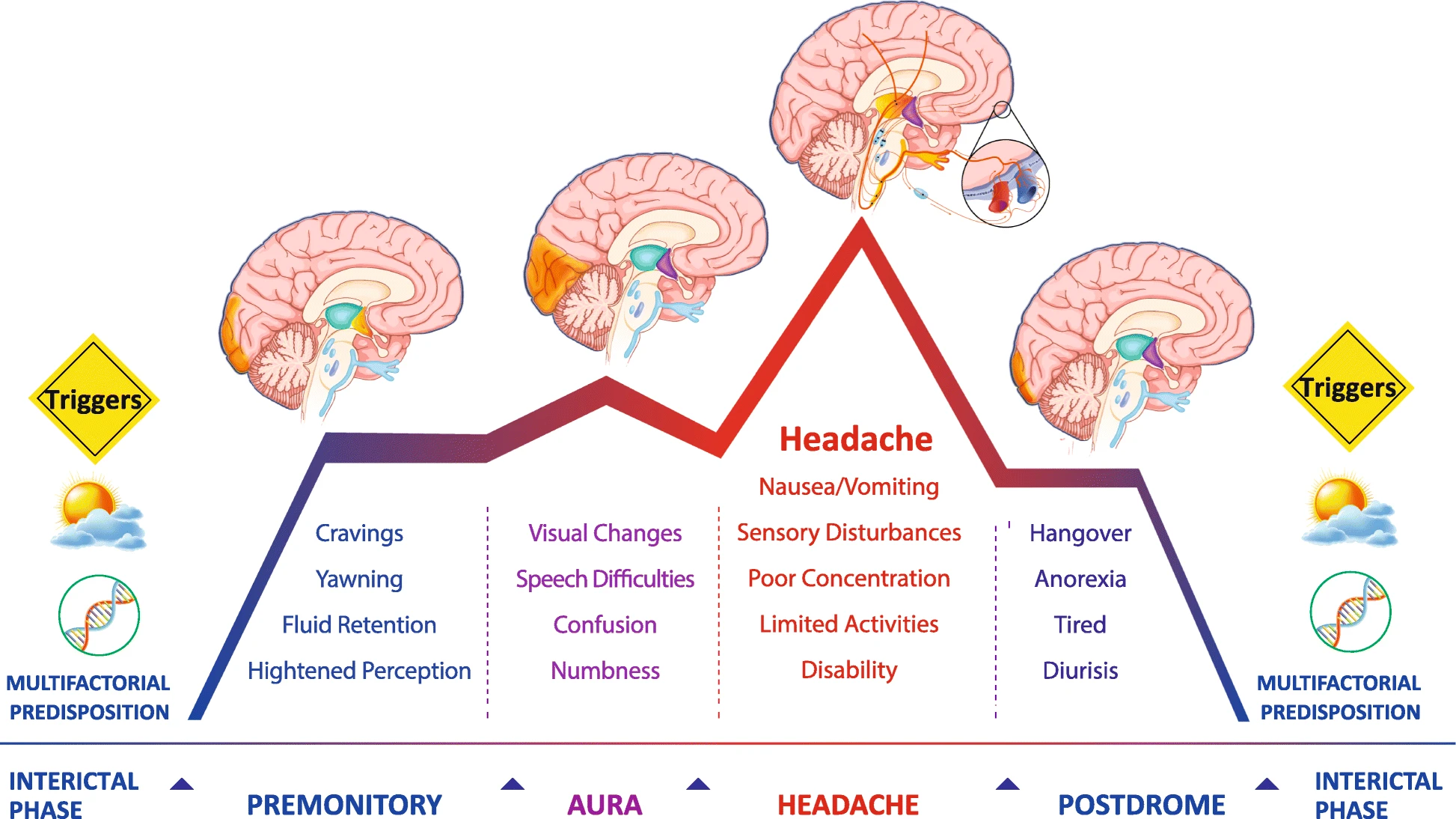
In some studies, it was found that gastric discomfort did not lead to headaches but actually gastric discomfort may mark the start of a migrainous headache and is called an aura. This all happens due to the effect of constriction of brain blood vessels and its effect on the corresponding autonomic area which controls our gastrointestinal system responses.

Different Phases of Migraine and Associated Symptoms
Different Phases of Migraine and Associated Symptoms (Andreou, A.P., Edvinsson, L. Mechanisms of migraine as a chronic evolutive condition. J Headache Pain 20, 117 (2019). https://doi.org/10.1186/s10194-019-1066-0)


Lifestyle or Dietary Factor Leading to Headaches


There are many stimulants that can be a reason for the start of migrainous headaches along with separately causing gastrointestinal symptoms. These include:
  • Stress
  • Not eating on time leading to hypoglycemia
  • Fatigue
  • Lack of sleep
  • Having foods that are known to stimulate an attack of migraines: Dark Chocolate, Cheese, Chinese food, Canned meat, and Alcohol.

Pathways for Headaches in GI disorders

While in another set of studies, for example, treatment of gastroesophageal reflux disease by giving proton pump inhibitors led to the resolution of headaches. In this set of studies, some of the hypotheses which have been proposed for this association are (1):
  • central sensitization and parasympathetic referred pain
  • serotonin pathways
  • autonomic nervous system dysfunction
  • systemic vasculopathy
  • food allergy.
However, still we don’t have an exact answer to how gastric and intestinal disorders can have an effect on the brain. It is an area still open to research to suggest a causal mechanism. But one thing is for sure that human body systems are closely knit together and are likely to have a bigger interplay between them than what we yet understand.

Check out These Top selling Books on Headaches 


Related Posts

History Taking in Neurosurgery : Headache

What are the pain sensitive intracranial structures? Brain itself is pain insensitive.  The following intracranial structures ar...


Hypertensive Pontine Hemorrhage

Contents Case Report Introduction Signs and symptoms Pathophysiology Diagnosis Treatment Hyperte...


References

1. T Noghani, Majid et al. “Gastrointestinal Headache; a Narrative Review.” Emergency (Tehran, Iran) vol. 4,4 (2016): 171-183.


2. Egilius L.H. Spierings (2002). Headache of Gastrointestinal Origin: Case Studies. , 42(3), 217–219. doi:10.1046/j.1526-4610.2002.02054.x 

3. Andreou, A.P., Edvinsson, L. Mechanisms of migraine as a chronic evolutive condition. J Headache Pain 20, 117 (2019). 


Contents

  1. Case Report
  2. Introduction
  3. Signs and symptoms
  4. Pathophysiology
  5. Diagnosis
  6. Treatment
  7. Hypertensive Pontine Hemorrhage Video

Case Report


  • 52yr old male
  • k/c/o HTN not on regular medication
  • Presented with sudden onset loss of consciousness


On Examination


  • GCS - E1VetM1
  • Pupils -B/L 1mm NRTL (Pin-Point Pupils)
  • Planters B/L mute


CT Scan
Hypertensive Pontine Hemorrhage CT Scan
Hypertensive Pontine Hemorrhage


  • Seen in long standing poorly-controlled chronic hypertension. 
  • It carries a very poor prognosis.
  • Pontine hemorrhage accounts for 5-10% of all hemorrhagic strokes (Jang et al 2011)


Signs and symptoms

  • "classic" pontine hematoma syndrome characterized by coma, quadriparesis, and eventual demise (Kushner et al 1985)
  • Clinical features of Pontine Hematoma are (Deng and Gaillard et al.)
    • Loss of consciousness (most common)
    • long tract signs including tetraparesis
    • cranial nerve palsies
    • seizures
    • Cheyne-Stokes respiration
  • CST does not play an essential role in recovery of independent walking and vestibulospinal tracts may not crucially affect recovery of independent walking in patients with pontine hemorrhage. In contrast, and intact CRP (corticoreticular pathway) or changes of the CRP integrity appear to be related to the recovery of gait function (Yeo et al 2020)

Pathophysiology

Hypertensive Pontine Hemorrhage
  • Due to rupture of penetrating arteries from the basilar artery extending into the pons 
  • These arteries are prone to lipohyalinosis as a result of poorly-controlled hypertension 
  • This makes the vessel wall prone to rupture. 
  • Larger paramedian perforators are more commonly the culprit vessels
Other Causes of Pontine Hemorrhage
Other causes of pontine hemorrhage include
  • Cavernoma
  • AV Malformation
  • Tumour bleed
  • Transtentorial herniation (Duret Hemorrhage)


Diagnosis

NCCT Brain is the investigation of choice. It shows:
  • Acute intraparenchymal hemorrhage within the pons
  • The hematoma frequently ruptures into the 4th ventricle 

Treatment

  • Poor prognosis
  • Large bleeds are almost universally fatal. 
  • Open surgical evacuation of the clot is usually not performed
  • In smaller hemorrhages, medical management and treatment of hydrocephalus with extraventricular drains may be life saving, 
  • Mortality ranges between 30% and 90% (Jang et al 2011)
  • Outcome depends on the volume of the bleed and initial GCS


Read Similar Posts

HISTORY TAKING IN NEUROSURGERY : HEADACHE


ACUTE SUBDURAL HEMATOMA WITH EXTRADURAL HEMATOMA: CT COMMA SIGN


ENDOVASCULAR TECHNIQUES FOR MANAGEMENT OF BRAIN ANEURYSMS: CONCEPTUAL ANIMATION VIDEOS

Neuro Urology and The Neurogenic Bladder

1. Neurogenic bladder refers to dysfunction of the urinary bladder due to disease of the central nervous system or peripheral nerves involved in the control of micturition . 
2. Non Neurogenic bladder refers to dysfunction of the urinary bladder due to dynamic disturbance of genitourinary system. 
 
Micturition Pathway
3. detrusor muscle of the bladder is innervated by parasympathetic neurons located in the S2-S4  column
 

The CT comma sign is a characteristic sign seen in head trauma. It is the presence of concurrent epidural and subdural hematomas, which gives the characteristic appearance of this sign as a "comma" shape.

The CT comma sign is a characteristic sign seen in head trauma. It is the presence of concurrent epidural and subdural hematomas, which gives the characteristic appearance of this sign as a "comma" shape.

The CT comma sign is a characteristic sign seen in head trauma. It is the presence of concurrent epidural and subdural hematomas, which gives the characteristic appearance of this sign as a "comma" shape.
The CT comma sign is a characteristic sign seen in head trauma. 
It is the presence of concurrent epidural and subdural hematomas, 
which gives the characteristic appearance of this sign as a "comma" shape.
           

70/M 

Chief Complaints: Numbness Lt Arm, 1 Episode of Partial seizure,Lt grip weakness since today morning. 

H/o Fall 3 days back. 

K/C/O HTN/DM/Post CABG 1994 with implanted defibrillator device (CRTD)/ Post Mechanical Thrombectomy for Rt MCA territory Infarct (March2020) on Ecospirin and T. APIXABAN(last dose today). 

Kn chronic  smoker and alcoholic. 

O/E 

E4V5M6
Pupils B/L 2mm RTL . 
Power Lt side 4/5. 
Left Hand grip 70%.

Since the patient was having multiple comorbidities with deranged coagulation and the GCS was 15, it was decided to manage the patient conservatively. Ecospirin and APIXABAN were stopped after consulting with a cardiologist and neurologist. 

#neuroradiology#neuroscience
#neurosurgeon #brainmri #braincat 

Reference


    MRI Spectroscopy : Neurosurgery Notes

    1. MR spectroscopy provides a measure of brain chemistry.
    2. The most common nuclei that are used are 
      • 1H (proton)
      • 23Na (sodium)
      • 31P (phosphorus). 
      • Proton spectroscopy is easier to perform and provides much higher signal-to-noise than either sodium or phosphorus.
    3. MRS can be performed within 10-15 minutes and can be added on to conventional MR imaging protocols. 
    4. It can be used to serially monitor biochemical changes in tumors, stroke, epilepsy, metabolic disorders, infections, and neurodegenerative diseases. 
    5. They require interpretation and should always be correlated with the MR images before making a final diagnosis.
    Hypothalamus Neuroanatomy/ Neurosurgery Notes


     Video Link:Neurosurgery written board crash course - hypothalamus 


    1. hypothalamus as the name suggests is directly underneath the thalamus and it's directly above the pituitary 
    2. borders of the hypothalamus can be drawn by a triangle 
      • line between the anterior commissure and a posterior commissure and it's called the ACPC line
      • line between the anterior commissure and the optic chiasm roughly boarded by the lamina terminalis 
      • line between the chiasm and the posterior commissure 


Q1. The “Nissl substance” represents which organelle of neuron?
A. Golgi complex
B. Nucleolus
C. Rough endoplasmic reticulum
D. Mitochondria

Ans: The cytoplasm of a neuron shows the presence of a granular material that stains intensely with basic dyes called Nissl substance (also called Nissl bodies or granules) . These bodies are rough endoplasmic reticulum

Q2. Which of the following provides myelin sheath to the axons of the CNS?
A. Astrocytes
B. Oligodendrocytes
C. Microglia
D. Ependymocytes

Ans: Oligodendrocytes form myelin sheath in CNS. Schwann cells form myelin sheath in PNS

Q3. The perivascular foot of the “blood–brain barrier” is an extension from the:
A. Oligodendrocyte
B. Ependymocyte
C. Astrocyte
D. Microglia

Ans: Astrocytes form the perivascular feet around a capillary. Astrocytes act as insulators, nourish the neurons, help form blood-brain barrier.

Q4. Sensation of pain is detected by:A. Mechanoreceptor
B. Chemoreceptor
C. Nociceptor
D. Thermoreceptor


Ans: Nociceptor

Q5. The cerebral aqueduct is developed from the cavity of:A. Rhombencephalon
B. Mesencephalon
C. Telencephalon
D. Diencephalon


Ans: The cavity of each telencephalic vesicle becomes the lateral ventricle. The cavity of  iencephalon (along with the central part of the telencephalon) becomes the third ventricle. The cavity of the mesencephalon remains narrow, and forms the cerebral aqueduct (aqueduct of Sylvius). The cavity of the rhombencephalon forms the fourth ventricle. Its continuation in the spinal cord is the
central canal


Q6. The failure of closure of the cranial end of neural tube gives rise to:A. Anencephaly
B. Hydrocephalus
C. Microcephaly
D. Meningomyelocoele


Ans: The neural tube remains open in the region of the brain because of nonclosure of the anterior neuropore. This results in anencephaly. Brain tissue, which is exposed, degenerates

Q7. By which week of intrauterine life does the neural tube close?
A. Fourth
B. Fifth
C. Sixth
D. Seventh


Ans: At 4 weeks the neural tube is normally completely closed (Ref: https://embryology.med.unsw.edu.au/embryology/index.php/Timeline_human_development#Neural)

Q8. The cervical flexure of the neural tube occurs:A. Between the forebrain and midbrain
B. In the midbrain
C. Between hindbrain and spinal cord
D. In the hindbrain
 

Ans: The cervical flexure lies at the junction of the rhombencephalon and the spinal cord  


Q9:Rabies virus, from the site of bite, travels along nerves by 
A. Forward Axoplasmic Flow
B: Reverse Axoplasmic flow
C: Dendritic Flow
D: Along the Endoneurium of nerve fibres


Ans: Rabies virus, from the site of bite, travels along nerves by reverse axoplasmic flow.

Q10:Polio virus is also transported from the gastrointestinal tract through :


A. Forward Axoplasmic Flow
B: Reverse Axoplasmic flow
C: Dendritic Flow
D: Along the Endoneurium of nerve fibres

Ans: Polio virus is also transported from the gastrointestinal tract through reverse axoplasmic flow.


Q11:Tetanus bacteria, in contrast, travels from the site of infection to the brain by:
A. Forward Axoplasmic Flow
B: Reverse Axoplasmic flow
C: Dendritic Flow
D: Along the Endoneurium of nerve fibres

Ans: Tetanus bacteria travels from the site of infection to the brain along the endoneurium of nerve fibres
  
Reference:

Dorsal Root Ganglion Anatomy
Dorsal Root Ganglion (Malanowski et al)


Lesion in Dorsal root ganglion of a spinal nerve in the neck is most likely to lead to what type of loss?

A. Sensory
B. Motor
C. Sympathetic
D. Parasympathetic
E. All of the above

Explanation:


The functions of dorsal root ganglia are all associated with the perception of sensations. These include:


  • Nociception
  • Perception of mechanical stimulus upon compression
  • First processing center of the sensory information
  • Role in reflex action


The clinical significance of dorsal root ganglion includes the treatment of chronic pain according to the gate-therapy or via complete resection of the ganglion. The ganglion also serves as a source or reservoir of several viruses such as herpes simplex virus.


References:

https://human-memory.net/dorsal-root-ganglion/

Malinowski M.N., Bremer N.J., Kim C.H. (2019) Dorsal Root Ganglion Stimulation. In: Abd-Elsayed A. (eds) Pain. Springer, Cham. https://doi.org/10.1007/978-3-319-99124-5_189


History Taking in Neurosurgery : Headache

What are the pain sensitive intracranial structures?

Brain itself is pain insensitive. 

The following intracranial structures are pain-sensitive:
  1. Meningeal arteries
  2. Proximal portions of the cerebral arteries
  3. Dura at the base of the brain
  4. Venous sinuses
  5. Cranial nerves 5, 7, 9, and 10, and cervical nerves 1, 2, and 3

What are the mechanisms causing headaches?

  • Distortion or traction of Dura, Venous Sinuses or Blood vessels: 

    • Drainage of CSF in erect posture causes headache, secondary to traction on the venous sinuses when the brain sinks toward the tentorium as it loses CSF flotation
    • Intracranial mass distorts the dura or the arteries at the base of the brain 
    • Distortion due to raised ICP 

  • Distension of a vessel

    • Distension of extracranial and occasionally intracranial arteries is thought to be the cause of pain in migraine (activate the trigeminal nerve terminals in the vessel wall)

  • Inflammation

    • Inflammation in the subarachnoid space can result in headache. Inflammation can be caused by infection, hemorrhage, or chemical irritation
    • Inflammation of vessel wall by autoimmune process. eg Giant cell arteritis

  • Referral of Pain

    • Lesions above the tentorium - referred pain in trigeminal nerve distribution (the forehead or behind the eye) - because the dura in this region is supplied by the trigeminal nerve
    • Lesions in the posterior fossa
      • referred pain in the ear and the back of the head - because this part of the dura is supplied by cranial nerves 9 and 10 and the upper three cervical roots
      • refer pain to orbit -  termination of orbital (ophthalmic division) pain nerve fibers in the lowest part of the spinal nucleus of the trigeminal nerve, which also receive termination of the upper cervical pain afferent nerve fibers 
      • referred pain to ear - Irritation of cranial nerves 7, 9, and 10 - because the ear has cutaneous supply from each of these nerves as well as cranial nerve 5.

What are the types of Headaches?

For the purpose of history taking, headaches can be divided into two types:
  • Primary- no identifiable cause on examination or investigation and diagnosis is based on  recognizing a pattern, e.g.
    • Migraine
    • Cluster headache
    • Tension-type headache
  • Secondary - definite identifiable cause on examination or investigation, e.g. 
    • Brain tumors
    • Meningitis
    • Sub-arachnoid hemorrhage 
  • 90% - primary headaches, less than 10% are secondary headaches (Rasmussen 1991)

What are the headache "Red Flags"?

  • Worst Headache ever
  • New onset Headache
  • Onset after age of 50 yrs
  • Change in pattern of headache
  • Worsening headache
  • Sudden onset during exertion, sneezing, coughing
  • Headache with postural variation
  • Headache in setting of malignancy or HIV
  • Headache associated with Neurological symptoms or signs
  • Associated with systemic symptoms - fever, weight loss and chronic cough

What history to take in a patient presenting with Headache?


Duration
Exact duration as reported by patient
NEW onset headache or something that has been there since before and has worsened now

Onset ( sudden/ gradual )
Acute onset, severe, first and worst headache, the common possibilities are sub-arachnoid hemorrhage, vascular dissection, pituitary apoplexy
Gradual onset – migraine(mins to days), SDH, GCA(days to months)

Severity
verbal rating scale from 0 to 10

Character
Pulsatile or throbbing or hammering (Raised ICP Headaches/ Migraine)
Dull featureless pain (Tension type headache)
Boring sharp- cluster HA

Time of occurrence
Raised ICP headaches - More in morning, May waken the patient at an early hour

Frequency
Increase in frequency - red flag

Distribution
Frontal or Holocranial - Raised ICP
Band like - tension headaache


Aggravating factors
Exertion, coughing, sneezing, stooping, and straining at stool
Changes in posture (increases in supine - ↑ICP , increases in upright – low CSF pressure headache)

Relieving factors
Improvement on lying flat - low pressure headache

Associated features
Blurring of Vision - Papilloedema
Diplopia, the commonest cause of which is abducens nerve paresis
Nausea & vomiting – migraine, ↑ICP
Neck stiffness – meningeal process
Changes in consciousness
Focal neurological symptoms


References


Diagram of Pupillary Light Reflex Pathway
Pupillary Light Reflex Pathway #neurosurgeon #neuro #neurosurgery #neurological #Pupil #neuroscience #neurology #neurochirurgie #lightreflex #pupillary #Lightreflexpathway https://ift.tt/33tMQAo via WSF Instagram

Anatomy of Basal Ganglia - Axial View

Structures generally included in the basal ganglia are the caudate, putamen, and globus pallidus in the cerebrum, the substantia nigra in the midbrain, and the subthalamic nucleus in the diencephalon.

Basal ganglia's primary function is likely to control and regulate activities of the motor and premotor cortical areas so that voluntary movements can be performed smoothly.

Dysfunction results in a wide range of neurological conditions including disorders of behaviour control and movement.

Those of behaviour include Tourette syndrome, obsessive–compulsive disorder, and addiction.

Movement disorders include, most notably Parkinson's disease, which involves degeneration of the dopamine-producing cells in the substantia nigra, Huntington's disease, which primarily involves damage to the striatum, dystonia, and hemiballismus


 #neurosurgeon #neuro #neurosurgery #neurological #basalganglia #neuroscience #neurology #neurochirurgie #neuroradiology #BGBleed #caudate #caudatenucleus https://ift.tt/33tMQAo via WSF Instagram

Topics